AI & digital pathology
AI & Digital Pathology
An AI pipeline that reads immune infiltration and tissue architecture from standard slides, built over four years with an industry partner.
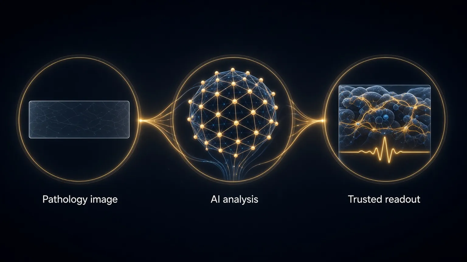
Digital pathology produces images faster than any pathologist can read them. The bottleneck is turning an image into a measurement a clinician can act on, reliably, on any scanner, in any laboratory. This program builds that layer, in a four-year collaboration with an Australian photonics start-up and with clinical collaborators in colorectal cancer.
The pipeline segments cells and tissue compartments, quantifies immune infiltration and collagen architecture, and combines those features into a stratification score. It runs on brightfield slides from ordinary scanners, with interpretable features a pathologist can inspect, and with the validation scaffolding, held-out cohorts and cross-site checks, that decides whether a readout deserves trust. Seed funding and a national healthtech competition placing supported its early development.
The strength is the combination: biology-led feature design, engineering discipline in validation, and a route to the clinic that does not depend on specialist hardware. Findings to date are retrospective; prospective validation is the next step.