Spatial biology & imaging
Spatial Imaging & Tissue Architecture
Label-free imaging of tissue architecture on the microscopes pathology labs already own, developed with an Australian photonics start-up.
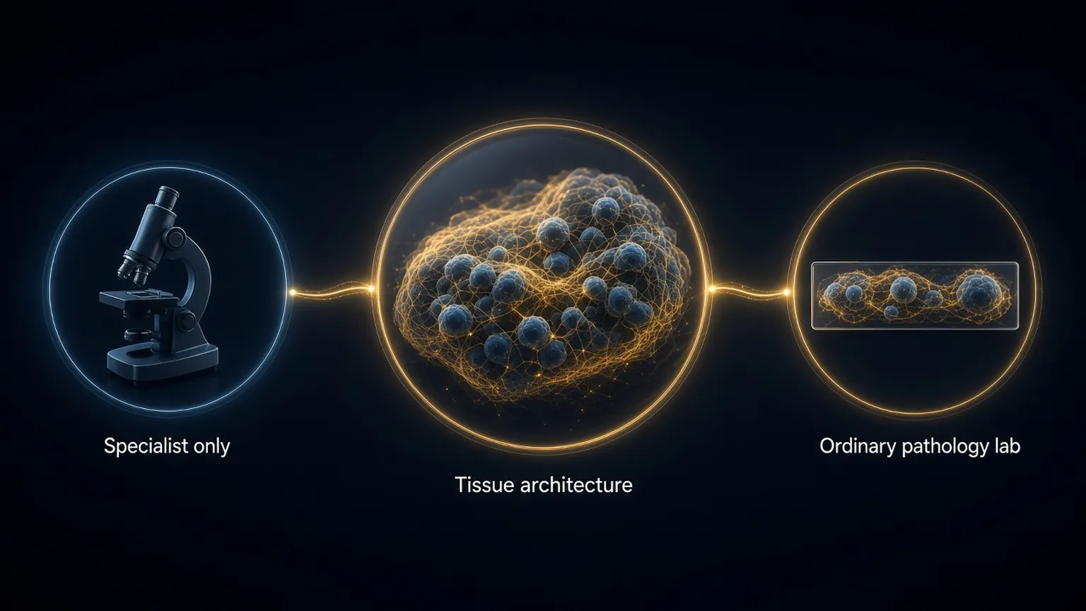
The collagen architecture around a tumour carries prognostic information, but reading it has needed second-harmonic or multiphoton microscopy that few centres own. For four years I have worked with an Australian photonics start-up on a different route: nano-structured microscope slides that let a standard brightfield microscope show collagen and cell structure without stains.
My part is the biology and the validation: defining what the images must show, benchmarking them against gold-standard instruments on matched tissue, and building the analysis that turns the optical signal into measurements of fibre alignment, density, and organisation around tumour cells. The work spans several solid tumour types and feeds directly into the AI and digital pathology program.
The strength is access. Structural information that used to need a specialist facility becomes something any pathology laboratory can capture on its existing equipment, which is the precondition for using it in routine care.